Using a microscope for the first time can be a profound experience. Suddenly, you can observe the world on a scale that is otherwise invisible to the naked eye. Imagine being one of the first researchers to combine multiple optical lenses and resolving organisms or structures that were previously just theoretical: Bacteria, cellular structures, and so on. The effect would have been sensational – and with good reason. The onset of microscopy pioneered new schools of thought while compound microscopes gradually became a staple instrument in virtually every research facility on the planet. It is odd to think that this ground-breaking success all hinged on a few lenses - and has since advanced to one of the most innovative, technical fields in the world of science. Thanks to microscopy and the development of microscopy filters, we now experience and understand life in a way we never thought possible.
Microscopy Filters 101
Lenses and filters are a staple of any instrument concerned with the physico-optical properties of sample materials. They are critical for observing the way that light of specific wavelengths reflects, scatters, diffracts off a surface, or is absorbed and emitted by it. This is how you can visualize the minute spatial and structural properties of samples under test. When you are observing weak emission signals like fluorescence or phosphorescence, you must use highly specialized microscopy filters.
The Main Components of Microscopy Filter Sets
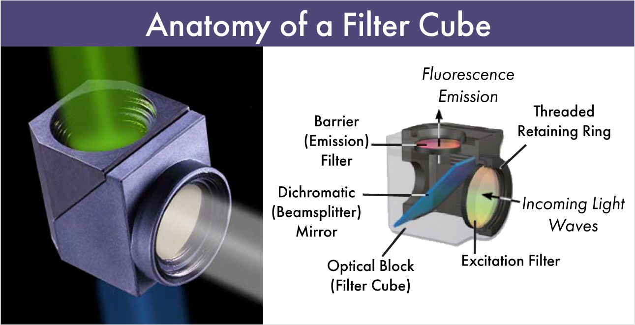
- An excitation filter, which is integrated into a cube, slider or wheel positioned in the light excitation path – between the light source and the objective lens;
- A dichroic mirror, which performs the dual function of reflecting excitation light to the sample while transmitting emission signals through to the eyepiece or detector;
- An emission filter, which is positioned between the objective lens and the eyepiece to screen out signals that are irrelevant to sample fluorescence (i.e. stray excitation light).
The Principles of Microscopy Filters
Though there is no universal workflow to explain how all fluorescence microscopy filters work, there is a set of basic principles that are worth bearing in mind when it comes to selecting your filter set.
Fluorescence microscopy concerns relatively weak emission signals only emitted by certain materials. These photo-luminescent compounds absorb light of specific wavelengths and emit photons when excited. Fluorophores typically stop fluorescing as soon as the excitation source is removed. The purpose of the fluorescence set is to direct the excitation wavelengths toward the sample and the emission wavelengths toward the detector. These are often housed in "cubes" with the excitation filter pointing towards the light source and the emission filter pointing towards the detector with the dichroic in between. The dichroic filter enables the entire setup. It serves two functions- as a steering mirror to reflect the excitation light into the sample and as a wavelength selector- passing emission wavelengths through towards the detector.

Once the filtered excitation light has passed through the excitation filter, it reflects off the dichroic mirror at a 45° angle and excites the fluorophores in the sample. The dichroic mirror is vital as it can reflect over 90% of the excitation light while transmitting over 90% of the emission light.
The longer wave fluorescence signals pass through the emission filter which performs a similar function to the exciter in that it passes a range of wavelengths while thoroughly blocking the excitation wavelengths. What is left produces the high contrast image of fluorescently-stained molecules on a black background.
Microscopy Filters from Omega Optical
As we have explained throughout this article, the performance of each of these components is based purely on their ability to selectively attenuate, transmit, or reflect light of specific wavelengths or within a given spectral region. You can typically distinguish between microscopy filters as either short- or long-pass filters, which governs which end of the electromagnetic spectrum their waveband is based in. The filter set you employ depends on your application and your labeling parameters. If you would like to experiment with fluorescence sets and fluorophores, try our Curvomatic.
Compare Fluorescence Sets and Fluorophores
Or, if you would like to speak with a member of the Omega Optical team about specific microscopy filters for your application, simply contact a member of the team today.
